CBCT · Guide · UK 2026
Crown first, implant second: a UL6 case from Dr Kunal Shah
I spend a lot of my working life talking about CBCT. Fields of view, voxel sizes, software keys. I'll admit I can get carried away with a spec sheet. So it's good to step back now and then and look at what all of that actually does for a patient. This case from Dr Kunal Shah at Leo Dental in London does exactly that.
The patient
A 40-year-old man came in with discomfort when chewing on his upper left side. His upper left first molar (UL6) was missing, food was packing into the space, and the area was hard to keep clean.
He had a very good reason to sort it. More than ten years earlier he'd lost his lower left second premolar, and the first molar behind it drifted into the gap. He didn't want that to happen again, or for the opposing tooth to over-erupt. Dr Shah went through every option with him: no treatment, a removable prosthesis, a fixed bridge, or an implant. He already had an implant in his upper right, so he knew the process, and he chose an implant.
Where CBCT came in
The anatomy to respect here was the maxillary sinus, which sat close to the planned site. That's exactly the sort of thing you want to see properly before anyone picks up a drill.
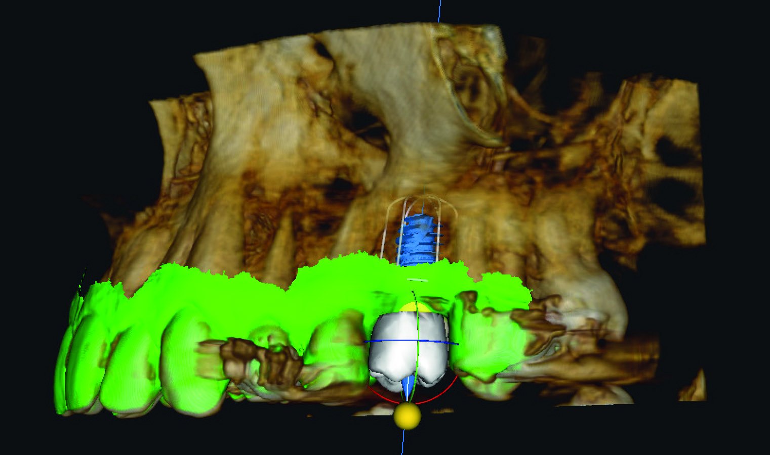
Dr Shah took an 8 x 9 volume on a Carestream Dental CBCT, then imported an intraoral scan and matched it to the CBCT in CS 3D Imaging Premium. That gave him the bone, the soft tissue and the teeth in one place.
He then planned crown first. The ideal crown was set on screen before any implant went in, and the software's AI-assisted auto implant planning proposed an implant position based on that crown, the surrounding anatomy and the available bone.
The bit I like most is what came next. Dr Shah checked the proposal against what matters in the posterior maxilla: the distance to the sinus, the bucco-palatal width of the ridge and the angulation. The software proposes. The clinician decides. As the case write-up puts it, AI proposals are an adjunct, not a replacement for clinical judgement.
The outcome
The plan could have been turned into a surgical guide, but in this case Dr Shah chose a conventional freehand approach with the digital plan as his reference. He placed an Anthogyr Axiom X3, chosen for its suitability in the posterior maxilla, and the implant was placed successfully in the UL6 region.
Dr Shah reports reduced planning time and simpler data handling, with the CBCT and intraoral scan matched in one place. Bone dimensions, crown position and the distance to the sinus were all visible on one plan.
You can read the full case, with all the images, here: AI-Assisted Implant Planning: Crown First, Implant Second.
Thank you to Dr Kunal Shah BDS PGCE, Principal Dentist at Leo Dental, for sharing it.
Thinking about CBCT for your own practice?
A case like this starts with the right scan. Every machine we supply takes an excellent scan, but the right one for you depends on your caseload, your room, your software and the dentistry you want to keep in-house. The field of view you choose decides which cases stay with you and which get referred out.
We've put together a guide that explains what the specs mean in real clinical work, and our team is happy to talk it through with you. We scan more than 3,000 patients a year on these machines in our own Yorkshire clinic, so the advice comes from daily use rather than a brochure.
Find out which CBCT is right for your practice
Pete